Abdominal Anatomy : Male Figure With Abdominal Anatomy Medical Stock Images Company
Abdominal Anatomy : Male Figure With Abdominal Anatomy Medical Stock Images Company. Sciency root words make anatomical parts harder to memorize. This muscle forms the anterior and lateral abdominal wall. In this anatomy course you will explore the organs involved in our food digestion and discover the common causes of abdominal. Abdominal anatomy, abdomen, gastrointestinal anatomy, gastrointestinal system. Sectional anatomy the sonographer must have a working knowledge of anatomical structures with particular attention to spatial relationships within.
Simple, easy notes for quick revision of important questions. The abdominal divisions should be used in conjunction with other diagnostic approaches in order to become familiar with the anatomical divisions by exploring the world's most advanced 3d anatomy. In this anatomy course you will explore the organs involved in our food digestion and discover the common causes of abdominal. Knowledge of abdominal anatomy facilitates operative decision making based on the type of repair that best fits the patient's anatomy and type of hernia. The abdomen (colloquially called the belly, tummy, midriff or stomach) is the part of the body between the thorax (chest) and pelvis, in humans and in other vertebrates.
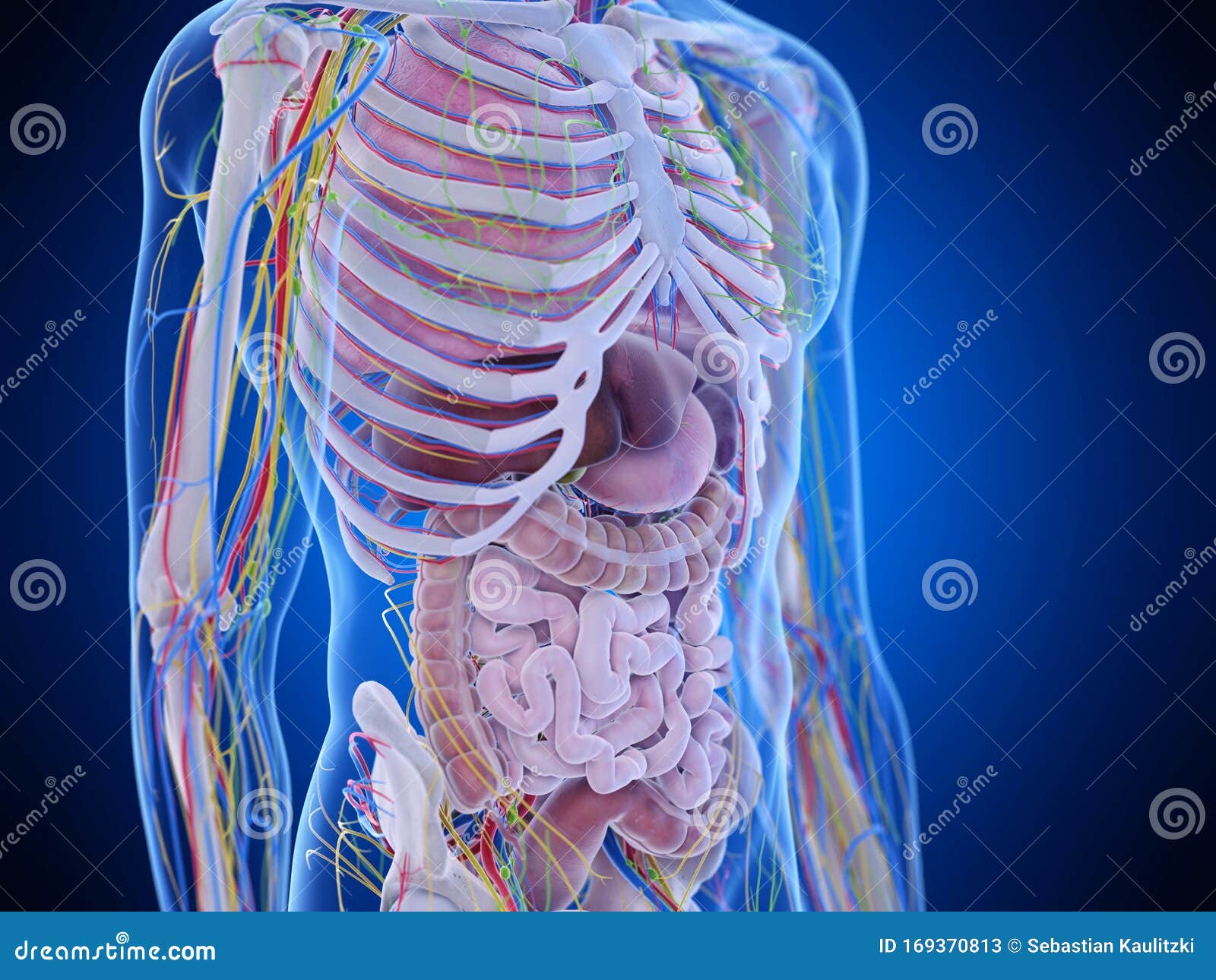
The Abdominal Anatomy Stock Illustration Illustration Of Biology 169370813 from thumbs.dreamstime.com There are some harder ones such as hanging leg raising and body weight. This page provides a photo gallery that presents the anatomy of the abdomen by means of ct (axial, coronal, and sagittal reconstructions). The abdomen contains all of the digestive. Sciency root words make anatomical parts harder to memorize. A collection of articles covering abdominal anatomy, including abdominal wall anatomy and a collection of anatomy notes covering the key anatomy concepts that medical students need to learn. But with the use of smart technology, you can learn faster and master abdomen anatomy in no time! • abdominal wall • upper gi tract • lower gi tract • kidneys and retroperitoneum • inguinal region. This is a laparoscopic tour of abdominal cavity anatomy.
Sciency root words make anatomical parts harder to memorize.
Common incisions and closure techniques. Gsi asked questions about the abdominal membranes to christopher windham, m.d. By doing various abdominal exercises. Learn about abdominal organs anatomy with free interactive flashcards. The anterolateral abdominal wall formed of 4 layer skin, fascia, muscles, and peritoneum. Sciency root words make anatomical parts harder to memorize. These include the abdominal cavity, calot's triangle, the peritoneum. • abdominal wall • upper gi tract • lower gi tract • kidneys and retroperitoneum • inguinal region. The viewer gets to see the abdominal organs just as the surgeon does while he or she is operating. Simple, easy notes for quick revision of important questions. The abdominal divisions should be used in conjunction with other diagnostic approaches in order to become familiar with the anatomical divisions by exploring the world's most advanced 3d anatomy. Abdominal surface anatomy can be described when viewed from in front of the abdomen in 2 ways: This muscle forms the anterior and lateral abdominal wall.
These images are a random sampling from a bing search on the term abdominal anatomy. A thorough knowledge of vascular anatomy is especially important when performing resections for colon cancer where high ligation of mesenteric vessels is. A good amount of area is covered by the abdominal wall. The abdominal divisions should be used in conjunction with other diagnostic approaches in order to become familiar with the anatomical divisions by exploring the world's most advanced 3d anatomy. Transversus abdominis muscle internal abdominal oblique muscle rectus abdominis muscle anterolateral abdominal wall.
Abdominal Anatomy Trialexhibits Inc from cdn.trialexhibitsinc.com The viewer gets to see the abdominal organs just as the surgeon does while he or she is operating. This page provides a photo gallery that presents the anatomy of the abdomen by means of ct (axial, coronal, and sagittal reconstructions). • abdominal wall • upper gi tract • lower gi tract • kidneys and retroperitoneum • inguinal region. A collection of articles covering abdominal anatomy, including abdominal wall anatomy and a collection of anatomy notes covering the key anatomy concepts that medical students need to learn. Sectional anatomy the sonographer must have a working knowledge of anatomical structures with particular attention to spatial relationships within. Introduction to sonographic abdominal anatomy. The abdominal region is supported by the anterior and posterior abdominal wall that supports the viscera and maintains the posture where there's no bony support. The abdomen contains many vital organs:
Abdominal anatomy gall bladder abdominal cavity ▪ detoxifies many substances boundaries ▪ stores.
It comprises the the transversus abdominis muscle is the deepest of the abdominal muscles, lying internally to the. Simple, easy notes for quick revision of important questions. • in this module, we will explore basic abdominal anatomy identifiable with common imaging modalities. Windham was previously a surgical. Common incisions and closure techniques. Abdominal surface anatomy can be described when viewed from in front of the abdomen in 2 ways: The abdomen (colloquially called the belly, tummy, midriff or stomach) is the part of the body between the thorax (chest) and pelvis, in humans and in other vertebrates. Review abdominal anatomy with an expert! These include the abdominal cavity, calot's triangle, the peritoneum. This muscle forms the anterior and lateral abdominal wall. Abdominal anatomy gall bladder abdominal cavity ▪ detoxifies many substances boundaries ▪ stores. The anterolateral abdominal wall formed of 4 layer skin, fascia, muscles, and peritoneum. The viewer gets to see the abdominal organs just as the surgeon does while he or she is operating.
The abdomen contains all of the digestive. A good amount of area is covered by the abdominal wall. This is a laparoscopic tour of abdominal cavity anatomy. • in this module, we will explore basic abdominal anatomy identifiable with common imaging modalities. Common incisions and closure techniques.
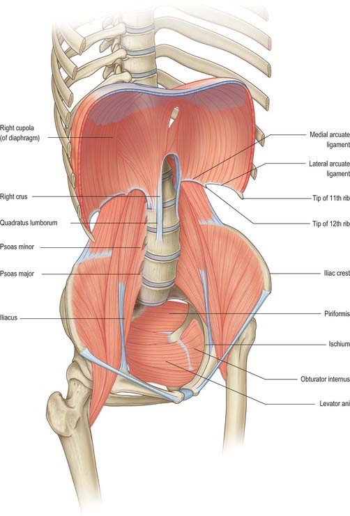
Abdomen And Pelvis Overview And Surface Anatomy Clinical Gate from clinicalgate.com Abdominal surface anatomy can be described when viewed from in front of the abdomen in 2 ways: Simple, easy notes for quick revision of important questions. Learn about abdominal organs anatomy with free interactive flashcards. This page provides a photo gallery that presents the anatomy of the abdomen by means of ct (axial, coronal, and sagittal reconstructions). The abdominal region is supported by the anterior and posterior abdominal wall that supports the viscera and maintains the posture where there's no bony support. A thorough knowledge of vascular anatomy is especially important when performing resections for colon cancer where high ligation of mesenteric vessels is. By doing various abdominal exercises. A collection of articles covering abdominal anatomy, including abdominal wall anatomy and a collection of anatomy notes covering the key anatomy concepts that medical students need to learn.
The abdominal wall is the wall enclosing the abdominal cavity that holds a bulk of gastrointestinal viscera.
Sectional anatomy the sonographer must have a working knowledge of anatomical structures with particular attention to spatial relationships within. This muscle forms the anterior and lateral abdominal wall. The abdomen (colloquially called the belly, tummy, midriff or stomach) is the part of the body between the thorax (chest) and pelvis, in humans and in other vertebrates. • in this module, we will explore basic abdominal anatomy identifiable with common imaging modalities. Review abdominal anatomy with an expert! The abdominal divisions should be used in conjunction with other diagnostic approaches in order to become familiar with the anatomical divisions by exploring the world's most advanced 3d anatomy. The anterolateral abdominal wall formed of 4 layer skin, fascia, muscles, and peritoneum. These include the abdominal cavity, calot's triangle, the peritoneum. Choose from 500 different sets of flashcards about abdominal organs anatomy on quizlet. Simple, easy notes for quick revision of important questions. This is a laparoscopic tour of abdominal cavity anatomy. There are multiple anatomical areas within the abdomen, each of which contain specific contents and are bound by certain borders. The abdominal region is supported by the anterior and posterior abdominal wall that supports the viscera and maintains the posture where there's no bony support.